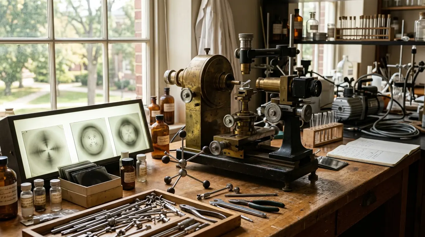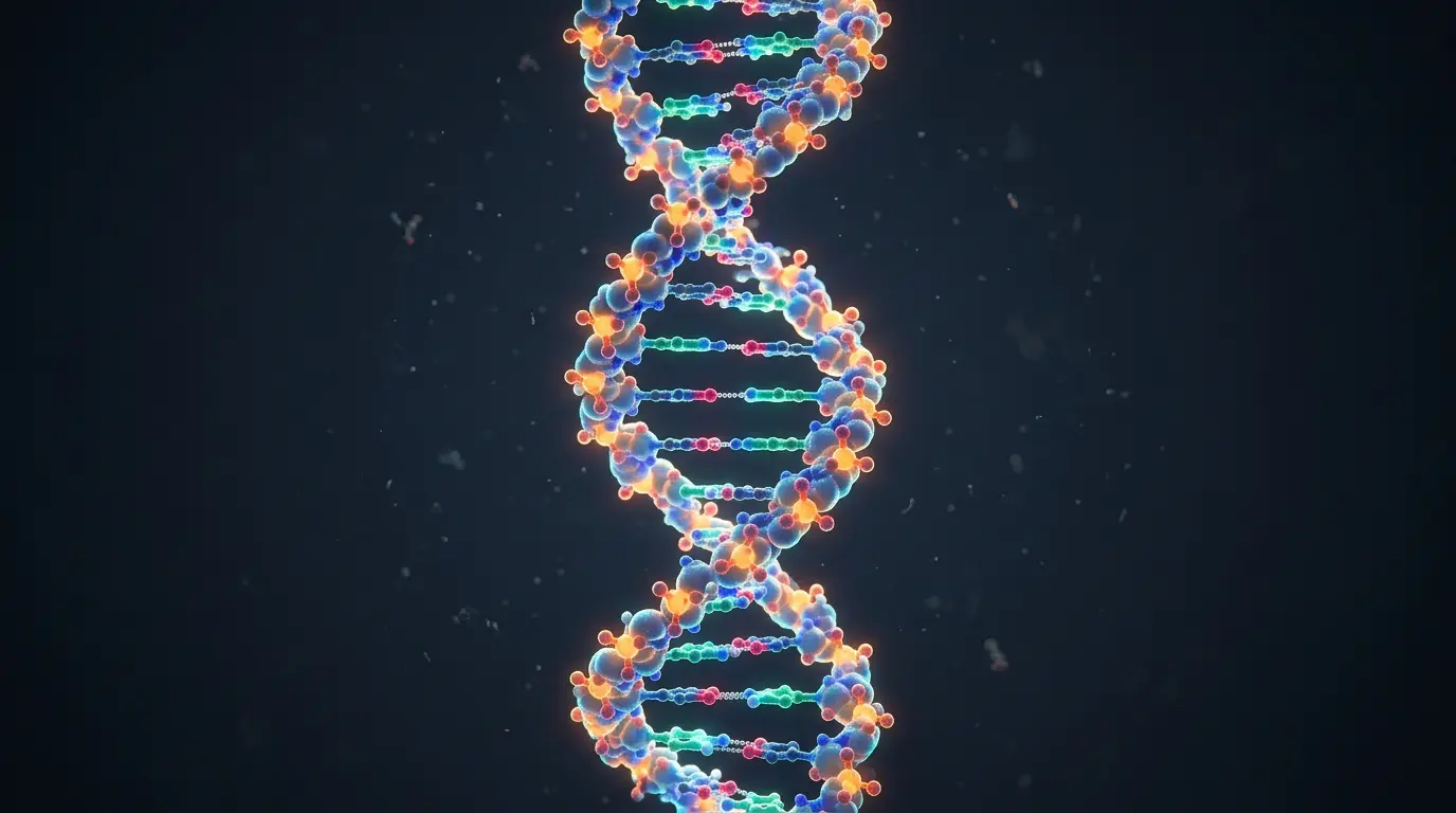
In the mid-20th century, discovering how living cells store and replicate genetic information stood as biology's ultimate frontier. Although deoxyribonucleic acid (DNA) was recognized as the hereditary material, its molecular architecture remained completely unknown. That profound puzzle was unraveled in the spring of 1953 through the vital intersection of meticulous X-ray crystallographic data and insightful three-dimensional model building.
Photo 51: Rosalind Franklin's Crystallographic Breakthrough
At King's College London, physical chemist and X-ray crystallographer Rosalind Franklin, working with graduate student Raymond Gosling, investigated the physical properties of DNA fibers. In May 1952, they produced Photo 51, a remarkably clear X-ray diffraction pattern of paracrystalline B-form DNA.
The unmistakable "X" diffraction pattern in Photo 51 provided unequivocal physical evidence of a helical geometry. Through rigorous mathematical calculations, Franklin deduced fundamental structural parameters: a diameter of 2 nanometers, a repeat period of 3.4 nanometers per full helical turn (spanning 10 base pairs), and a hydrophilic sugar-phosphate backbone located externally, shielding the hydrophobic nitrogenous bases tucked inside.
Watson and Crick's Structural Model Building
Concurrently, at the Cavendish Laboratory at Cambridge University, James Watson and Francis Crick pursued the solution through physical scale-model construction. In late January 1953, Maurice Wilkins showed Photo 51 to Watson without Franklin's knowledge or consent. Furthermore, an MRC committee report containing Franklin's data on monoclinic C2 space-group symmetry allowed Crick to deduce that the two polynucleotide chains must run in opposite, antiparallel directions.
A critical piece of the puzzle fell into place when physical chemist Jerry Donohue corrected textbook errors regarding nitrogenous base tautomers, pointing out that thymine and guanine exist predominantly in the keto rather than enol form. This insight enabled Watson to identify the exact hydrogen-bonding geometry:
- Adenine (A) pairs specifically with Thymine (T) via two hydrogen bonds.
- Guanine (G) pairs specifically with Cytosine (C) via three hydrogen bonds.
This pairing explained Erwin Chargaff's 1950 empirical finding that the ratios of A to T and G to C are always equal in cellular DNA.
Publication in Nature and Historical Legacy
On April 25, 1953, the journal Nature published three consecutive papers: Watson and Crick's theoretical structure, Wilkins and colleagues' optical data, and Franklin and Gosling's diffraction analysis. Watson and Crick famously closed their seminal paper with a classic understatement:
"It has not escaped our notice that the specific pairing we have postulated immediately suggests a possible copying mechanism for the genetic material."
In 1962, the Nobel Prize in Physiology or Medicine was awarded jointly to Watson, Crick, and Wilkins. Tragically, Rosalind Franklin had died of ovarian cancer in 1958 at age 37, and the Nobel Foundation strictly prohibits posthumous awards. Today, the global scientific community unequivocally honors Franklin's indispensable role as a primary discoverer of the double helix.